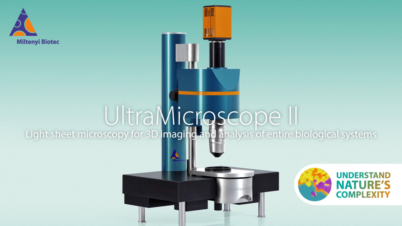
UltraMicroscope II Light Sheet Microscope
Manufacturer: Miltenyi Biotec
SKU: UltraMicroscope II
Description
The UltraMicroscope II is a light sheet fluorescence microscope configured for generating detailed three-dimensional images of large biological samples, including whole organs, embryos, and cleared tissues. It provides two primary optical configurations: a Zoom Body Module, which offers a magnification range from 1.26x to 12.6x and is suited for multi-user environments due to its ease of use, and a Super Plan Module, which delivers higher resolution with a magnification range from 0.66x to 30x. The system supports both unidirectional and bidirectional illumination schemes. The light source can be either a laser module with up to five specific laser lines (405 nm, 488 nm, 561 nm, 639 nm, 785 nm) or a supercontinuum white light laser covering a broad spectrum from 460 to 800 nanometers. A notable feature is the automated compensation for refractive index, which allows the microscope to maintain image quality when used with various tissue clearing protocols and imaging media. This flexibility facilitates the visualization of entire biological systems, from an overview down to cellular resolution, making it a tool for research in fields requiring large-volume 3D imaging.
Specifications
| Item | UltraMicroscope II Light Sheet Microscope |
| Company | Miltenyi Biotec |
| Catalog Number | UltraMicroscope II |
| Quantity | EA |
| Type | Cell Imaging System, Digital, Fluorescence Imaging, Light Sheet Microscope |
| Total Magnification | 1.26x to 12.6x (Zoom body config)0.66x to 30x (Super plan config) |
| Illumination | Uni- and bidirectional |
| Light Source | Laser module (max. 5 laser lines): 405 nm, 488 nm, 561 nm, 639 nm, 785 nm OR Supercontinuum white light laser: 460 to 800 nm |
| Applications | Fluorescence imaging of large (mm-sized) specimens, Detection and localization of single cells in entire biological systems, Large-scale 3D visualization of structural features and networks under normal and pathological conditions, Time-lapse LSFM |
| Objectives | Zoom body configuration: 2 x NA 0.5 (diagonal FOV 1.7 to 17.6 mm) Super Plan configuration: 1.1 x NA 0.1, 4 x NA 0.35, 12 x NA 0.53 (diagonal FOV up to 33 mm with 1.1x) Total magnification : 1.26x to 12.6x (Zoom body config), 0.66x to 30x (Super plan config) |
| Sample Type | Oganoids, rodent organs, mouse embryos, mouse brain, larvae, tumors, tissue sections, biopsies etc. Sample size up to approx. 1 cm |
| Detector(s) | Camera-based (sCMOS) 5.5 Megapixel with 100 fps and 60% quantum efficiency OR 4.2 Megapixel with 100 fps and 82% quantum efficiency |
Vendors & Pricing
No vendors listed yet
Vendor pricing will appear here once available.