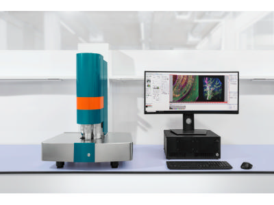
UltraMicroscope Blaze™ Light Sheet Microscope
Manufacturer: Miltenyi Biotec
SKU: Blaze
Description
The UltraMicroscope Blaze is an automated light sheet fluorescence microscope engineered for high-speed, high-resolution three-dimensional imaging of large, cleared tissue specimens. Its design aims to streamline the workflow for imaging multiple samples, such as organoids, rodent organs, or entire small animal models, with applications in neuroscience, immuno-oncology, and developmental biology. A key feature is the LightSpeed mode, which accelerates data acquisition significantly compared to conventional light sheet methods, enabling tasks like capturing a full 3D image of an entire mouse brain in under three minutes. The system incorporates automation for critical functions including objective exchange, autofocus, and chromatic correction. Imaging is performed using camera-based sCMOS detectors, with options for a 4.2 or 5.5-megapixel camera. Illumination is provided by either a laser beam combiner supporting up to five laser lines (405, 488, 561, 639, 785 nm) or a supercontinuum white light laser. The microscope includes application-specific sample holders, some with a clamping mechanism, designed for imaging multiple samples sequentially in a batch mode. Image processing is handled by the dedicated MACS iQ View software, which offers tools for destriping, denoising, deconvolution, and stitching of large volumetric datasets.
Specifications
| Item | UltraMicroscope Blaze™ Light Sheet Microscope |
| Company | Miltenyi Biotec |
| Catalog Number | Blaze |
| Quantity | EA |
| Applications | Cleared tissue imaging, 3D imaging, wide range of applications in neuroscience, immuno-oncology, and developmental biology, suitable for academia, industry, and pre-clinical studies. |
| Sample Holder | Application-specific sample holders designed for imaging multiple samples (e.g., 8 mouse brains, 48 organoids) seamlessly, incorporating a correlated template mode for hands-free sequential imaging. |
| Detector(s) | Camera-based (sCMOS), 4.2 Megapixel with 100 fps and 82% quantum efficiency or 5.5 Megapixel with 100 fps and 60% quantum efficiency. |
| Light Source | Laser beam combiner (max. 5 laser lines with 405, 488, 561, 639, 785 nm, 50 mW–100 mW per diode) or supercontinuum white light laser (405–850 nm, 1–4 mW/nm). |
| Objectives | Zoom body configuration: 2x NA 0.5 (diagonal FOV 1.7–17.6 mm) Automated objective (AO) configuration: 1.1x NA 0.1, 4x NA 0.35, 12x NA 0.53 (diagonal FOV up to 33 mm with 1.1x) |
| Type | Light sheet microscope, fluorescence microscope. |
| Sample Type | Transparent and cleared 3D samples. multiple samples, or samples with a size of up 4.4 cm × 7.0 cm × 1.7 cm (x,y,z) and even bigger, like organoids, tissue sections, biopsies, tumors, whole organs, embryos (all e.g. rodent, human), or larvae. |
| Dimensions | (WxHxD) 67 x 91 x 52.5 cm |
Vendors & Pricing
No vendors listed yet
Vendor pricing will appear here once available.