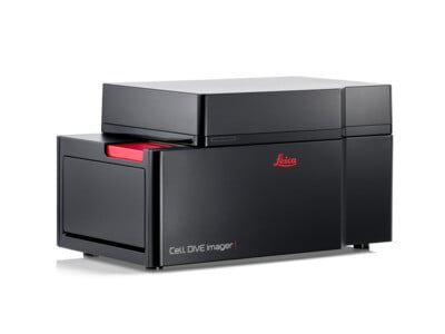
Cell DIVE Multiplexed Imager
Manufacturer: Leica Microsystems
Description
Leica Microsystems' Cell DIVE is an automated imaging platform for spatial proteomics, capable of visualizing more than 60 protein biomarkers on a single formalin-fixed, paraffin-embedded (FFPE) tissue section. The system employs a cyclic immunofluorescence method, where antibodies are applied, imaged, and then removed in an iterative process, allowing for high-plex analysis. It captures data at whole-tissue scale while resolving individual cells, enabling the study of cellular interactions, tissue microenvironments, and biomarker co-expression patterns. This technology is applicable in research areas such as cancer biology, immunology, and neuroscience, where understanding the spatial context of protein expression is critical. The platform supports the use of archival samples and is designed to generate quantitative, single-cell resolution data for complex spatial biology investigations.
Specifications
| Method | Multiplex immunofluorescence imaging of 60+ biomarkers |
| Scale | Whole tissue, single cell |
Vendors & Pricing
No vendors listed yet
Vendor pricing will appear here once available.